Filariasis
Filariasis is caused by several round, coiled and thread-like parasitic worms that belongs to the family filaridea. These parasites penetrate the skin either on their own or through the opening created by mosquito bites to reach the lymphatic system.
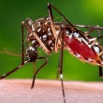
The disease is caused by the nematode worm, either Wuchereria bancrofti or Brugia malayi and is transmitted by mosquito species Culex quinquefasciatus and Mansonia annulifera/M.uniformis respectively.
The disease generally presents with the symptoms like swelling of legs, and hydrocele and can cause a raft of societal stigma.
Lymphatic Filariasis (LF) is also commonly known as elephantiasis. It is a disfiguring and disabling disease, which is generally seen in childhood. In the early stages, there are either no symptoms or non-specific symptoms. The lymphatic system is damaged. This stage can last for several years. Infected persons sustain the transmission of the disease. The long term physical consequences are painful swollen limbs (lymphoedema or elephantiasis). Hydrocele in males is also common in endemic areas.
Symptoms
Edema with thickening of the skin and underlying tissues is the classical symptom of filarasis.
It usually affects the lower extremities. However, the arms, vulva, breasts and scrotum (causing hydrocele formation) can also be affected.The edema in the extremities, breast or genital area can result in the part becoming several times its normal size and is due to blockage of the vessels of the lymphatic system. The incubation period of filaria is 10-14 days.
Other symptoms include:
Skin rashes
Hyper or hypo pigmented macules
River blindness (caused by Onchocerca volvulus)
Causes
Most cases of filaria are caused by the parasite known as Wuchereria bancrofti. The Culex, Aedes or Anopheles mosquitoes transmit the disease. Another parasite called Brugia malayi that causes filariasis is transmitted by the vector Mansonia and Anopheles mosquitoes.
When an infected mosquito bites a healthy person, the larvae called microfilariae move into the lymphatics and lymph nodes. Here, they develop into adult worms and may persist for years.
The adult parasite, in turn, produces more microfilariae. These microfilariae circulate in the peripheral blood usually in the night, and are sucked by the mosquitoes during a bite. The same cycle is then repeated in another healthy individual.
Diagnosis
Blood sample:
The microfilariae that cause lymphatic filariasis circulate in the blood at night (called nocturnal periodicity). Blood collection should be done at night to coincide with the appearance of the microfilariae, and a thick smear should be made and stained with Giemsa or hematoxylin and eosin. For increased sensitivity, concentration techniques can be used.
Serological examination:
Serologic techniques provide an alternative to microscopic detection of microfilariae for the diagnosis of lymphatic filariasis. Patients with active filarial infection typically have elevated levels of antifilarial IgG4 in the blood and these can be detected using routine assays.
For further diagnosis, consult your physician.
Treatments
Maintenance of Good hygiene of the affected part prevents the worsening of the lymphedema and secondary bacterial skin infections.
The affected limb should be kept elevated and regular exercises should be done to improve the lymph flow.
The recommended regimen for treatment through mass drug administration (MDA) is a single dose of two medicines given together - albendazole (400 mg) plus either ivermectin (150-200 mcg/kg) in areas where onchocerciasis (river blindness) is also endemic or diethylcarbamazine citrate (DEC) (6 mg/kg) in areas where onchocerciasis is not endemic. These medicines clear microfilariae from the bloodstream.
While medicines are available to treat filaria, the gross swelling of the leg makes a person look noticeable and ugly. Hence, it is better to protect from the bites of filaria mosquitoes.
For more queries, you should consult your doctor.
References:
WHO
CDC